Synchronous Oligometastatic Non-Small Cell Lung Cancer and Isolated Renal Cell

publicité
Open Access Case
Report
DOI: 10.7759/cureus.366
Synchronous Oligometastatic Non-Small
Cell Lung Cancer and Isolated Renal Cell
Carcinoma: A Case Report and Literature
Review
Timothy K. Nguyen 1 , Alexander V. Louie 2
1. Radiation Oncology, London Health Sciences Centre 2. Department of Radiation Oncology, London
Regional Cancer Program, Western University, London, Ontario, Canada
 Corresponding author: Timothy K. Nguyen, [email protected]
Disclosures can be found in Additional Information at the end of the article
Abstract
A 58-year-old gentleman presenting with a progressive headache, visual disturbance,
decreased appetite, and weight loss was found to have a localized clear cell carcinoma of the
kidney and synchronous Stage IV non-small cell lung cancer with a solitary brain metastasis.
This case illustrates the challenges in distinguishing between primary and metastatic disease in
a patient with both renal cell carcinoma and lung cancer. We highlight the uncertainties in the
diagnosis and management of this unique clinical scenario and the potential implications on
prognosis.
Categories: Oncology, Radiation Oncology
Keywords: synchronous, non-small-cell lung cancer, renal cell carcinoma, metastatic, metastasis,
oligometastasis, oligometastatic, concomitant, simultaneous, metastasectomy
Introduction
Received 10/01/2015
Review began 10/02/2015
Review ended 10/06/2015
Published 10/27/2015
© Copyright 2015
Nguyen et al. This is an open access
article distributed under the terms of
the Creative Commons Attribution
License CC-BY 3.0., which permits
unrestricted use, distribution, and
reproduction in any medium,
provided the original author and
source are credited.
The oligometastatic state is a clinical paradigm whereby cancer patients with limited
metastases (i.e., 1 to 5) that are amenable to aggressive management through resection or
ablative therapy may survive beyond expectations [1-2]. At the time of diagnosis, non-small cell
lung cancer (NSCLC) and renal cell carcinoma (RCC) present with metastatic disease in
approximately 56% and 30% of cases, respectively [3]. Renal metastases have been reported in
16-23% of metastatic NSCLC cases while, for metastatic RCC, the lungs are the most common
site of metastasis representing up to 45% of cases [4-5]. In situations with radiographic
evidence of both pulmonary and renal tumors, the diagnostic challenge becomes differentiating
between primary neoplasms and metastatic disease. We report a case of oligometastatic NSCLC
with a solitary brain metastasis in the setting of a synchronous primary renal neoplasm.
Informed patient consent was obtained prior to the drafting and submission of this report.
Case Presentation
Initial assessment
We present a case of a 58-year-old, right-handed gentleman with a medical history of Type II
diabetes, obstructive sleep apnea, gastroesophageal reflux disease, asthma, gout, anxiety, and
depression. He was an ex-smoker with a 40 pack-year smoking history. His oncologic family
history was significant for his father dying from bladder cancer.
How to cite this article
Nguyen T K, Louie A V (October 27, 2015) Synchronous Oligometastatic Non-Small Cell Lung Cancer and
Isolated Renal Cell Carcinoma: A Case Report and Literature Review. Cureus 7(10): e366. DOI
10.7759/cureus.366
In the spring of 2014, the patient initially presented to his local emergency room with
progressive frontotemporal headaches, a right visual field deficit, decreased appetite, weight
loss, and a fall without sustaining traumatic injuries. On a review of systems, no respiratory,
genitourinary, or additional neurological symptoms were described aside from increased
daytime urinary frequency.
Physical examination revealed an obese gentleman (BMI of 41) with normal vital signs. The
patient was alert, oriented to person, place and time, and ambulatory with a normal gait.
Cranial nerve exam revealed right visual field deficits. Auscultation of the lungs revealed clear,
equal air entry bilaterally without any adventitious sounds appreciated. No masses,
organomegaly, or pelvic lymphadenopathy were appreciated on the abdominal exam, but this
was limited due to his large body habitus. Digital rectal exam revealed a small, smooth, firm
prostate without rectal masses.
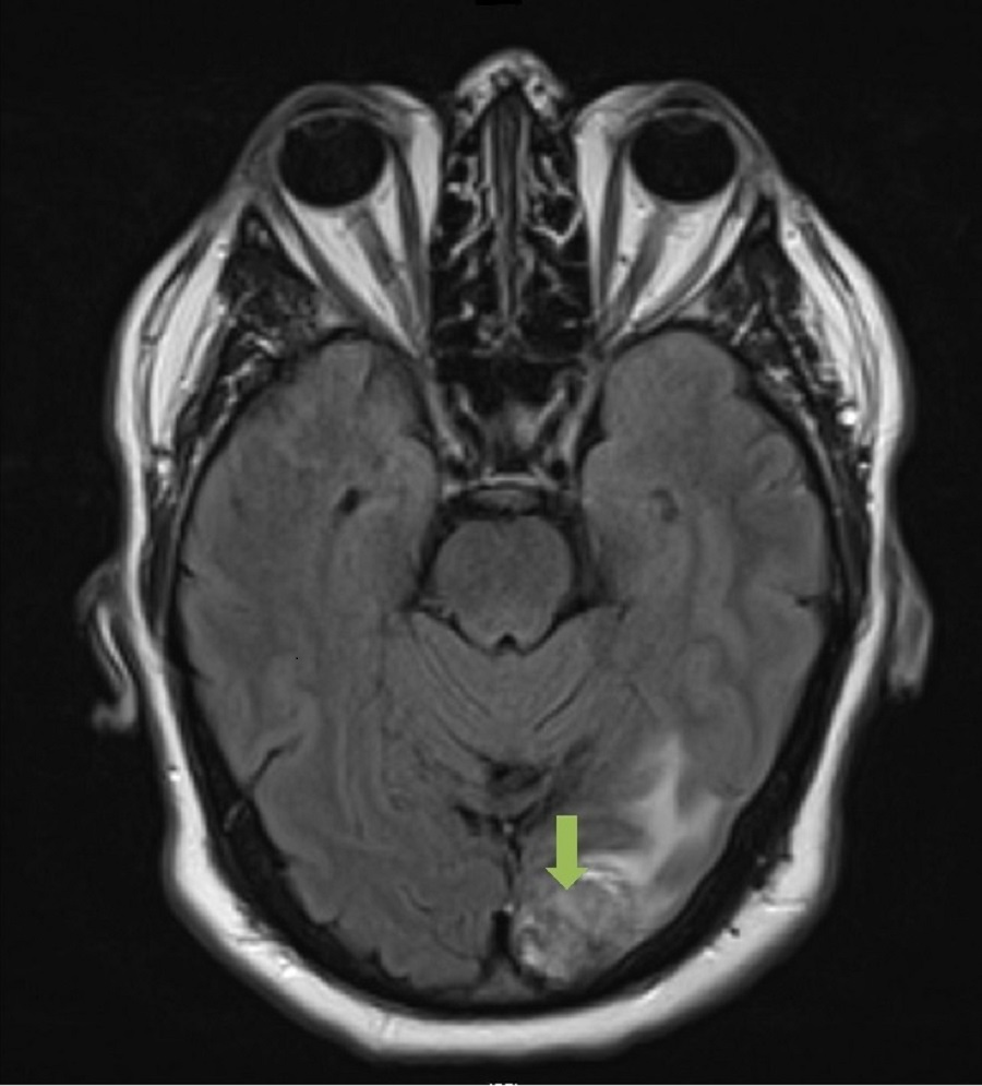
A CT and MRI of the head was completed with the latter revealing a heterogeneous mass in the
left occipital lobe with solid and cystic components measuring 2.9 cm. There was associated
cerebral edema with a 0.2 cm right-sided midline shift and no hydrocephalus identified (Figure
1).
FIGURE 1: Preoperative MRI of the Head
2015 Nguyen et al. Cureus 7(10): e366. DOI 10.7759/cureus.366
2 of 9
FIGURE 1: Preoperative MRI of the Head
Axial slice. T2-weighted image that demonstrates a solitary 2.9 cm left occipital lobe
adenocarincoma, metastatic from a lung primary.
Initial management
The leading differential diagnosis at the time was a primary brain malignancy, and accordingly,
referral to neurosurgery at the same tertiary hospital was arranged. The patient subsequently
underwent left occipital craniotomy and resection of the mass within five weeks of the initial
referral. Pathology returned as metastatic adenocarcinoma, suggestive of a lung primary as it
stained positive for cytokeratin 7 and TTF-1 and negative for cytokeratin 20. Adjuvant wholebrain radiotherapy was delivered to a dose of 30 Gy in 10 fractions, which was completed within
six weeks from the time of surgery. The start date of radiotherapy was delayed by a couple of
weeks as a result of a postoperative wound infection that was managed with oral antibiotics.
During this time, the patient had a staging CT scan in the community encompassing the thorax,
abdomen, and pelvis, which revealed a 5 cm left upper pole renal mass without other sites of
disease. This report was not available in the patient’s record; however, the dictated notes from
the medical team indicated that metastatic renal cell carcinoma was the provisional diagnosis.
Concomitant with the planning and delivery of whole brain radiotherapy, the patient was also
seen and assessed by the urology team at the same institution. Restaging CT imaging of the
thorax, abdomen, and pelvis was completed two weeks after completion of radiotherapy, which
revealed an exophytic solid renal mass arising from the upper pole of the left kidney measuring
4.9 cm. In addition, there were now two other areas of concern: a 3.1 cm heterogeneous
pancreatic mass containing cystic components and a 2.1 cm pulmonary nodule in the lower
lobe of the right lung. Both lesions, radiographically, were thought to be metastases from a
primary renal cell carcinoma. Considerations for management included cytoreductive surgery,
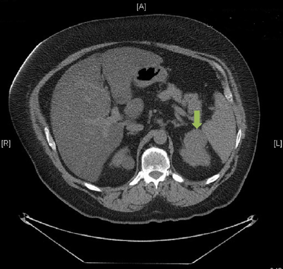
up-front Sunitinib, or enrolling the patient on a clinical trial (Figures 2-3).
2015 Nguyen et al. Cureus 7(10): e366. DOI 10.7759/cureus.366
3 of 9
FIGURE 2: Preoperative CT of the Abdomen
Axial slice. Contrast-enhanced image that demonstrates a 4.9 cm isolated clear cell renal cell
carcinoma involving the upper pole of the left kidney.
2015 Nguyen et al. Cureus 7(10): e366. DOI 10.7759/cureus.366
4 of 9
FIGURE 3: Preoperative CT of the Thorax
Axial slice. Contrast-enhanced image that demonstrates a 2.1 cm primary papillary
adenocarcinoma of the right lower lobe of the right lung.
However, before the patient could return to further discuss management options, an MRI scan
of the head, completed two months after finishing radiotherapy to assess treatment effect,
showed evidence of an intracranial abscess in the previous operative bed. Specifically, there
was an interval development of a 4.2 cm rim-enhancing fluid signal in the post-surgical bed.
Urgently, the patient was brought to the operating room for a left occipital craniectomy for
evacuation and resection of the abscess. The patient recovered well without any additional
complications, although did not wish to seek any further medical care at his local institution.
A second opinion
The patient’s primary care team in the community referred the patient to the multidisciplinary
genitourinary oncology team at our center for a second opinion regarding management of the
patient’s remaining disease. Following a review of the original imaging and pathology reports,
the possibility of metastatic lung cancer (given the immunohistochemistry of the resected brain
lesion favoring a lung primary) or synchronous primary malignancies was proposed. A PET-CT
scan was ordered, which re-demonstrated the pulmonary mass in the lower lobe of the right
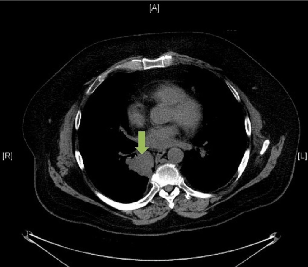
lung, now measuring 3.0 cm with an SUV uptake of 9. A 1 cm non-18-FDG-avid subcarinal
lymph node was also identified. The renal lesion measured 4.2 cm and was not 18-FDG-avid. A
nodule in the pancreatic tail was thought to be a benign process, given its cystic appearance,
stable size, and lack of 18-FDG uptake.
2015 Nguyen et al. Cureus 7(10): e366. DOI 10.7759/cureus.366
5 of 9
The case was also presented at our thoracic multidisciplinary team conference with two
recommendations provided. First, it was advised to complete the diagnostic workup, including
biopsies of both the lung and renal masses, in addition to sampling the mediastinal lymph
nodes. Second, depending on the results of the investigations, treatment should be considered
in accordance with the oligometastatic paradigm, which could involve sequential resection of
the pulmonary and renal tumors.
An ultrasound-guided biopsy of the kidney mass confirmed renal cell carcinoma (clear cell). The
sample stained positive for CKAE1/AE3 and vimentin, while staining negative for CK7, CK20,
and TTF-1. Through an endobronchial ultrasound (EBUS)-guided biopsy, tissue was obtained
from the right lower lobe mass and a paraesophageal (level 8) lymph node. The lymph node was
negative for malignancy while the biopsy from the mass showed the presence of neoplastic
cells. Given the limited sample, pathologists were unable to distinguish definitively between a
well-differentiated adenocarcinoma and a neuroendocrine tumor at that time. It was evident,
however, that the lung and kidney pathology were distinctly different.
Management
In accordance with recommendations from both our multidisciplinary teams, the patient
proceeded with radical intent treatment, first undergoing a video-assisted transthoracic
surgery (VATS) right lower lobe lobectomy within two months of initial assessment at our
center. Pathology revealed a unifocal tumor measuring 4.5 cm and was characterized as a Grade
2 papillary adenocarcinoma with negative margins. Eight lymph nodes were removed, and one
peribronchial lymph node was found to be positive for malignancy with extranodal extension.
Final lung pathologic staging was T2aN1.
Postoperatively, the patient met and discussed with the medical oncology team regarding the
benefits of adjuvant chemotherapy in the setting of N1 and oligometastatic disease. Given the
unclear benefit systemic therapy might provide for completely resected oligometastatic disease
in a patient with moderate comorbidities, adjuvant chemotherapy was ultimately declined by
the patient despite it being offered.
Restaging CT of the abdomen and pelvis was completed three months following VATS resection
and demonstrated that the kidney mass had grown in size to 6.4 cm since previous imaging. The
patient subsequently underwent a left partial nephrectomy three months later. Pathology
revealed a unifocal 6.0 cm tumor in the upper pole of the left kidney, which was resected with
negative margins. Histologically, the tumor was consistent with the previous biopsy,
characterized as a clear cell renal cell carcinoma. Regional lymph nodes were not sampled. The
final pathologic staging was pT1bNx, and no additional adjuvant treatment was felt to be
indicated.
The patient was seen in follow-up three months following resection of the renal mass, which
was 10 and 16 months from previous pulmonary and intracranial resections, respectively. He
recovered well without surgical complications and was found to have no clinical evidence to
suggest disease recurrence or progression. Repeat CT imaging of the head, thorax, abdomen,
and pelvis revealed no evidence of recurrent or residual disease. Moving forward, the patient
will continue with routine CT surveillance every three to six months.
Discussion
2015 Nguyen et al. Cureus 7(10): e366. DOI 10.7759/cureus.366
6 of 9
Synchronous primary malignancies are complex cases that present with diagnostic, prognostic,
and therapeutic uncertainties, particularly if there has already been metastatic spread from one
or both of the primary sites. We report on a case of oligometastatic adenocarcinoma of the lung
with a solitary brain metastasis that presents synchronously with a primary clear cell renal cell
carcinoma. The challenges surrounding this case stem from the propensity of lung cancer to
metastasize to the kidneys and brain, and likewise, the ability for kidney cancers to metastasize
to the lungs and brain. In such situations, histologic confirmation with immunohistochemistry
can help distinguish between metastatic and primary disease. Given the paucity of evidencebased standards to guide treatment decisions in these clinical scenarios, we propose that such
cases be discussed in the context of the relevant multidisciplinary tumor boards.
There have been a limited number of published cases reporting on the diagnostic and
therapeutic approach to synchronous primary pulmonary and renal neoplasms. Similar to our
case, a report from the Netherlands described a patient with a TTF-1 positive adenocarcinoma
of the right lower lobe of the lung invading into the thoracic vertebrae that presented
synchronously with an 18-FDG-negative lesion in the right kidney [6]. Initially, the case was
considered to be Stage IV NSCLC with the renal mass representing a distant metastasis and, as
such, was managed with an expectative approach. In the context of a second opinion and
multidisciplinary reevaluation, however, a provisional diagnosis of two synchronous primary
neoplasms was made. The lung cancer was managed with two cycles of neoadjuvant
chemotherapy (cisplatin and pemetrexed) followed by surgical resection. The kidney cancer was
managed with surveillance, given its stability in size, until it was later resected at the patient’s
request. The patient was disease-free at the six years following radical treatment, again
highlighting the difficulty and importance in differentiating between primary versus metastatic
disease.
Distinguishing between a renal metastasis of lung origin and a primary RCC (or vice versa) can
be a difficult task. The use of PET imaging can be useful in identifying metastatic deposits in the
kidneys, which tend to be 18-FDG-avid. However, a lack of hypermetabolic activity does not
rule out the possibility of a primary RCC. PET imaging has been described to have a low
sensitivity to detect primary renal malignancies, which was consistent with the experience of
Mazouz, et al. and with our own findings in the present case [6-7]. Although renal metastases
are typically bilateral and multi-focal, they can also present as a large, solitary metastasis in
breast, lung, and colorectal patients [8]. Thus, obtaining pathology is advised in patients
amenable for biopsy. In addition, only 1-3% of patients with metastatic RCC present with a
solitary metastasis [9]. When considered alongside additional supportive clinical evidence, this
point may steer clinicians away from a diagnosis of metastatic RCC in cases of synchronous
primary neoplasms with a solitary metastasis.
In a retrospective review of percutaneous core biopsies for renal masses, unnecessary surgery
was avoided in 30% of cases in which biopsies revealed benign tumors or non-surgical
metastatic disease [10]. Sanchez-Ortiz, et al. conducted a retrospective review of 100 patients
with a renal mass in the setting of a non-renal primary malignancy. Fifty-nine percent of renal
masses were malignant primary tumors, 19% were metastases (lung and lymphoma being the
most common primary sites), 12% were benign primary tumors, and 10% had non-diagnostic
biopsies. All cases of renal metastasis occurred in the setting of clinical progression or
radiographic evidence of other metastases from the non-renal primary cancer [11]. The authors
contend that for the majority of patients with a renal mass in the context of a clinically
localized non-renal malignancy, the renal tumor is unlikely to be metastatic disease. Once
pathology is obtained, immunohistochemical techniques can help direct diagnosis, with TTF-1
staining positive in approximately 76% of lung adenocarcinomas [12].
In the oligometastatic setting, there have been several publications that demonstrate a
prolonged survival when pursuing metastasectomies in intracranial, hepatic, and pulmonary
2015 Nguyen et al. Cureus 7(10): e366. DOI 10.7759/cureus.366
7 of 9
metastases [13-15]. However, randomized data to support a benefit in overall survival with
radical treatment for oligometastatic disease has only been demonstrated in the solitary brain
metastasis setting [16]. As the body of literature on the management of oligometastatic disease
continues to grow, heightened attention towards its implications on prognosis and treatment
will be vital in ensuring high-quality patient care.
Conclusions
In conclusion, we present a novel case of a 58-year-old gentleman presenting with a
synchronous localized renal cell carcinoma in the setting of oligometastatic NSCLC with a
solitary brain metastasis. In accordance with the oligometastatic paradigm, the patient is alive,
disease-free, and well 14 months from the initiation of his treatment. Management of
synchronous primary neoplasms, particularly in the oligometastatic setting, relies on a
thorough diagnostic workup and careful consideration of all possible clinical scenarios, which
should occur in the context of a multidisciplinary team approach.
Additional Information
Disclosures
Human subjects: Consent was obtained by all participants in this study.
References
1.
2.
3.
4.
5.
6.
7.
8.
9.
10.
11.
12.
Hellman S, Weichselbaum R: Oligometastases. J Clin Oncol. 1995, 13:8–10.
Palma DA, Salama J, Lo S, Senan S, Treasure T, Govindan R, Weichselbaum R: The
oligometastatic state – separating truth from wishful thinking. Nat Rev Clin Oncol. 2014,
11:549-57. 10.1038/nrclinonc.2014.96
Gay HA, Michalski JM: Cancer of the Kidney, Renal Pelvis and Ureter . Perez and Brady’s
Principles and Practice of Radiation Oncology 6th Edition. Halperin EC, Wazer DE, Perez CA,
Brady LW (ed): Lippincott Williams & Wilkins, Pennsylvania; 2013. 63:1245-1258.
Quint LE, Tummala S, Brisson LJ, Francis IR, Krupnick AS, Kazerooni EA, Iannettoni MD,
Whyte RI, Orringer MB: Distribution of distant metastases from newly diagnosed non-small
cell lung cancer. Ann Thorac Surg. 1996, 62:246–50. 10.1016/0003-4975(96)00220-2
Bianchi M, Sun M, Jeldres C, Shariat SF, Trinh QD, Briganti A, Tian Z, Schmitges J, Graefen M,
Perrotte P, Menon M, Montorsi F, Karakiewicz PI: Distribution of metastatic sites in renal cell
carcinoma: a population-based analysis. Ann Oncol. 2012, 23:973–80.
10.1093/annonc/mdr362
Mazouz A, Amaadour L, Souaf I, El Fatemi H, Amarti A, Erraisse MA, Oubelkacem E, Bouhafa
T, Tahiri Y, Tazi MF, Mellas S, Arifi S, Mellas N: Synchronous malignant renal mass in patient
with a lung cancer: case report and literature review. Pan Afr Med J. 2015, 20:22.
10.11604/pamj.2015.20.22.5541
Kang DE, White RL, Zuger JH, Sasser HC, Teigland CM: Clinical use of fluorodeoxyglucose F18 positron emission tomography for detection of renal cell carcinoma. J Urol. 2004,
171:1806–9. 10.1097/01.ju.0000120241.50061.e4
Choyke PL, White EM, Zeman RK, Jaffe MH, Clark LR: Renal metastases: clinicopathologic and
radiologic correlation. Radiology. 1987, 162:359–63. 10.1148/radiology.162.2.3797648
Montie JE, Stewart BH, Straffon RA, Banowsky LH, Hewitt CB, Montague DK: The role of
adjunctive nephrectomy in patients with metastatic renal cell carcinoma. J Urol. 1977,
117:272–75.
Lebret T, Poulain JE, Molinie V, Herve JM, Denoux Y, Guth A, Scherrer A, Botto H:
Percutaneous core biopsy for renal masses: indications, accuracy and results . J Urol. 2007,
178:1184–88. 10.1016/j.juro.2007.05.155
Sánchez-Ortiz RF, Madsen LT, Bermejo CE, Wen S, Shen Y, Swanson DA, Wood CG: A renal
mass in the setting of a nonrenal malignancy: when is a renal tumor biopsy appropriate?.
Cancer. 2004, 101:2195–201. 10.1002/cncr.20638
Harlamert HA, Mira J, Bejarano PA, Baughman RP, Miller MA, Whitsett JA, Yassin R: Thyroid
2015 Nguyen et al. Cureus 7(10): e366. DOI 10.7759/cureus.366
8 of 9
13.
14.
15.
16.
transcription factor-1 and cytokeratins 7 and 20 in pulmonary and breast carcinoma. Acta
Cytol. 1998, 42:1382–88. 10.1159/000332172
Patchell RA, Tibbs PA, Walsh JW, Dempsey RJ, Maruyama Y, Kryscio RJ, Markesbery WR,
Macdonald JS, Young B: A randomized trial of surgery in the treatment of single metastases
to the brain. N Engl J Med. 1990, 322:494–500. 10.1056/NEJM199002223220802
Morris EJ, Forman D, Thomas JD, Quirke P, Taylor EF, Fairley L, Cottier B, Poston G: Surgical
management and outcomes of colorectal cancer liver metastases. Br J Surg. 2010, 97:1110–18.
10.1002/bjs.7032
Weichselbaum RR, Hellman S: Oigometastases revisted. Nat Rev Clin Oncol. 2011, 8:378–82.
10.1038/nrclinonc.2011.44
Andrews DW, Scott CB, Sperduto PW, Flanders AE, Gaspar LE, Schell MC, Werner-Wasik M,
Demas W, Ryu J, Bahary JP, Souhami L, Rotman M, Mehta MP, Curran WJ Jr: Whole brain
radiation therapy with or without stereotactic radiosurgery boost for patients with one to
three brain metastases: phase III results of the RTOG 9508 randomised trial. Lancet. 2004,
363:1665–72. 10.1016/S0140-6736(04)16250-8
2015 Nguyen et al. Cureus 7(10): e366. DOI 10.7759/cureus.366
9 of 9
Téléchargement